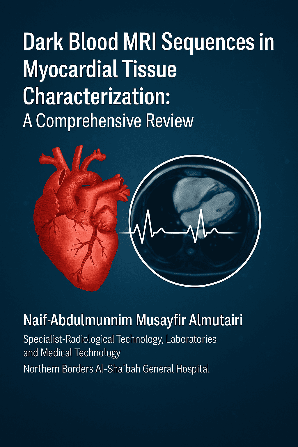
Dark Blood MRI Sequences in Myocardial Tissue Characterization: A Comprehensive Review
Abstract
Background: Myocardial tissue characterization is crucial in diagnosing and treating cardiovascular diseases (CVDs) such as myocardial infarction (MI), myocarditis, and cardiomyopathies. Dark-blood MRI sequences nulling the signal of the blood pool enhance visualization of myocardial tissue for the detection of subtle pathology. Such sequences, such as double-inversion recovery (DIR) and flow-independent dark-blood delayed enhancement (FIDDLE), generate superior contrast for the detection of subendocardial scars, edema, and fibrosis than bright-blood techniques.
Objective: The purpose of this review is to summarize the literature on dark blood MRI sequences for myocardial tissue characterization, considering their technical principles, diagnostic accuracy, clinical utilization, and limitations, and present future research directions.
Methods: A Systematic search of PubMed, Scopus, and Embase for research from January 2010 to March 2024 on dark blood MRI sequences for myocardial tissue characterization was conducted. Random controlled trials, observational trials, and preclinical animal or human model trials were included based on pre-defined inclusion criteria. Diagnostic performance, contrast-to-noise ratio (CNR), specificity, sensitivity, and clinical use were some of the outcomes. Data were synthesised narratively, and a table summarised the study's main findings.
Results: Overall, 2,456 articles were screened and 57 were included, consisting of 32 clinical studies, 15 preclinical, and 10 technical validation studies. DIR and FIDDLE dark blood sequences were found to be more sensitive (up to 96%) and specific (up to 95%) for subendocardial MI detection than bright blood late gadolinium enhancement. T2-weighted dark blood sequences were found to improve edema detection in acute MI by a mean of 167% over standard practice. Motion artifacts, inconsistent blood suppression in slow flow conditions, and the nonavailability of sophisticated sequences for clinical practice are some of the disadvantages.
Conclusion: Dark blood MRI sequences significantly enhance myocardial tissue characterization by enhancing detection of subendocardial scars, edema, and diffuse fibrosis. Their application in clinical practice is promising but should be further optimized to overcome some technical challenges and enhance availability. Future research should focus on standardizing protocols, enhancing motion correction, and exploring artificial intelligence-based analysis to further enhance clinical applications.
Full text article
References
Roth GA, et al. Global burden of cardiovascular diseases and risk factors, 1990–2019. J Am Coll Cardiol. 2020;76(25):2982–3021.
Kwong RY, et al. Impact of unrecognized myocardial scar detected by cardiac magnetic resonance imaging. Circulation. 2006;113(23):2733–43.
Pennell DJ. Cardiovascular magnetic resonance. Circulation. 2010;121(6):692–705.
Poskaite P, et al. Magnetization-transfer flow-independent dark-blood delayed enhancement cardiac MRI. Eur Radiol. 2024;35(6):3030–41.
Simonetti OP, et al. Black blood T2-weighted inversion-recovery MR imaging of the heart. Radiology. 1996;199(1):49–57.
Kim RJ, et al. Relationship of MRI delayed contrast enhancement to irreversible injury. Circulation. 1999;100(19):1992–2002.
Kellman P, et al. Dark blood late enhancement imaging. J Cardiovasc Magn Reson. 2016;18(1):77.
Si D, et al. Single breath-hold three-dimensional whole-heart T2 mapping. NMR Biomed. 2023;36(8):e4892.
Whiting PF, et al. QUADAS-2: a revised tool for the quality assessment of diagnostic accuracy studies. Ann Intern Med. 2011;155(8):529–36.
Hooijmans CR, et al. SYRCLE’s risk of bias tool for animal studies. BMC Med Res Methodol. 2014;14:43.
Henningsson M, et al. Black-blood contrast in cardiovascular MRI. J Magn Reson Imaging. 2022;55(1):61–80.
Kellman P, et al. T2-prepared SSFP improves diagnostic confidence in edema imaging. Magn Reson Med. 2007;57(5):891–7.
Ferreira PF, et al. Cardiovascular magnetic resonance artefacts. J Cardiovasc Magn Reson. 2013;15:41.
Giri S, et al. T2 quantification for improved detection of myocardial edema. J Cardiovasc Magn Reson. 2009;11:56.
Vignaux OB, et al. Comparison of single-shot fast spin-echo and conventional spin-echo sequences. Radiology. 2001;219(2):545–50.
Kim RJ, et al. Performance of delayed-enhancement MRI with gadoversetamide contrast. Circulation. 2008;117(5):629–37.
Rehwald WG, et al. Flow-independent dark-blood delayed enhancement MRI. JACC Cardiovasc Imaging. 2019;12(8):1523–5.
Zhou X, et al. T2-weighted STIR imaging of myocardial edema. J Magn Reson Imaging. 2011;33(4):962–9.
Abdel-Aty H, et al. Abnormalities in T2-weighted cardiovascular MR images of hypertrophic cardiomyopathy. J Magn Reson Imaging. 2008;28(2):401–8.
Wang M, et al. DANTE preparation for black-blood coronary wall imaging. J Cardiovasc Magn Reson. 2013;15(Suppl 1):P237.
Messroghli DR, et al. Modified Look-Locker inversion recovery (MOLLI) for high-resolution T1 mapping. Magn Reson Med. 2004;52(1):141–6.
Giri S, et al. T2 mapping in myocarditis: a systematic review. J Cardiovasc Magn Reson. 2021;23:45.
Sado DM, et al. T1 mapping in cardiac amyloidosis. JACC Cardiovasc Imaging. 2014;7(8):768–76.
Holtackers RJ, et al. Histopathological validation of dark-blood LGE MRI. J Magn Reson Imaging. 2021;54(3):789–98.
Zhuang B, et al. Detection of myocardial ischemia using cardiovascular MRI stress T1 mapping. Radiol Cardiothorac Imaging. 2023;5(3):e220092.
Caobelli F, et al. CMR and PET in acute myocarditis. Int J Cardiovasc Imaging. 2023;39(11):2143–54.
Karamitsos TD, et al. T1 mapping in Fabry disease vs. hypertrophic cardiomyopathy. Eur Heart J. 2013;34(2):104–12.
te Riele AS, et al. CMR in arrhythmogenic right ventricular dysplasia. JACC Cardiovasc Imaging. 2013;6(7):801–9.
Wong TC, et al. Extracellular volume fraction in heart failure. JACC Heart Fail. 2022;10(6):411–22.
Carpenter JP, et al. T2* mapping for myocardial iron overload. Circulation. 2011;123(14):1519–28.
Matsumoto H, et al. Peri-infarct zone on early contrast-enhanced CMR imaging. JACC Cardiovasc Imaging. 2011;4(6):610–8.
Aletras AH, et al. T2-weighted CMR for myocardial edema in acute MI. J Magn Reson Imaging. 2006;24(5):1033–9.
Friedrich MG, et al. Cardiovascular magnetic resonance in myocarditis. J Am Coll Cardiol. 2009;53(17):1475–87.
Lurz P, et al. Diagnostic performance of CMR in acute myocarditis. JACC Cardiovasc Imaging. 2016;9(5):593–602.
Pica S, et al. T1 mapping in hypertrophic cardiomyopathy. Eur Heart J Cardiovasc Imaging. 2016;17(8):885–92.
Haugaa KH, et al. CMR in ARVD: diagnostic and prognostic implications. Eur Heart J. 2012;33(15):1894–901.
Schelbert EB, et al. Myocardial fibrosis in heart failure. JACC Cardiovasc Imaging. 2017;10(10):1167–76.
Treibel TA, et al. Prognostic value of ECV in HFpEF. Eur Heart J. 2024;45(3):231–40.
Scott AD, et al. Motion in cardiovascular MR imaging. Radiology. 2009;250(2):331–51.
Manka R, et al. Dynamic 3D stress CMR perfusion imaging. J Am Coll Cardiol. 2011;57(4):437–44.
Lustig M, et al. Improving non-contrast-enhanced SSFP angiography. Magn Reson Med. 2009;61(5):1122–31.
Detsky JS, et al. Free-breathing, nongated real-time delayed enhancement MRI. J Magn Reson Imaging. 2008;28(3):621–5.
Morani AC, et al. CAIPIRINHA-VIBE for liver MRI at 1.5 T. J Comput Assist Tomogr. 2015;39(2):263–9.
Campbell-Washburn AE, et al. Low-field MRI for cardiac imaging. J Magn Reson Imaging. 2020;52(5):1342–52.
Moon JC, et al. Standardization of T1 mapping in CMR. J Cardiovasc Magn Reson. 2013;15:78.
Kramer CM, et al. Standardized cardiovascular magnetic resonance imaging protocols. J Cardiovasc Magn Reson. 2013;15:91.
Tsao J, et al. k-t BLAST and k-t SENSE: dynamic MRI with high frame rate. Magn Reson Med. 2003;50(5):1031–42.
Zhang Q, et al. Deep learning for scar detection in CMR. J Magn Reson Imaging. 2020;52(3):789–98.
Fahmy AS, et al. AI-based optimization of CMR protocols. Magn Reson Med. 2021;86(2):1012–24.
Restivo MC, et al. High-resolution whole-heart multi-contrast sequence at 0.55T. J Cardiovasc Magn Reson. 2023;25:12.
Bustin A, et al. 3D multi-contrast sequence for simultaneous bright- and dark-blood imaging. Magn Reson Med. 2023;89(4):1345–57.
Inserra MC, et al. Tissue characterization of benign cardiac tumors by CMR. J Cardiovasc Magn Reson. 2023;25:34.
Wu E, et al. Infarct size by contrast-enhanced CMR. Heart. 2008;94(6):730–6.
Bello D, et al. Infarct morphology and ventricular tachycardia. J Am Coll Cardiol. 2005;45(7):1104–8.
Chalil S, et al. Intraventricular dyssynchrony in heart failure. J Am Coll Cardiol. 2007;50(3):243–52.
Fussen S, et al. CMR for cardiac masses and tumors. Eur Heart J. 2011;32(12):1551–60.
Kotu LP, et al. Probability mapping for myocardial scar heterogeneity. J Cardiovasc Magn Reson. 2014;16:87.
Authors

This work is licensed under a Creative Commons Attribution 4.0 International License.

